
Guest lecture on zebrafish intestinal cell research
Emeritus Prof Dr Paul Thomas Matsudaira from the Centre for Bioimaging Sciences of the National University of Singapore (NUS) delivered a session of guest lecture to UTAR students and staff on 21 July 2023 at Tan Sri Lee Loy Seng Lecture Theatre (EDK 1), UTAR Kampar Campus. The topic of the lecture was “Zebrafish stem cells in the primitive intestinal crypts”.
The main research areas of Prof Paul are the mechanobiology of cells and tissues on viscoelastic surfaces, nanoscale dynamics, and bioimaging sciences. He studied chemistry and later biology at the University of Washington, US. In 1981, he obtained a Doctor of Philosophy in biology from Dartmouth College, US. He furthered his studies at the Max Planck Institute for Biophysical Chemistry in Germany and the Laboratory of Molecular Biology, UK. His academic career began at the Whitehead Institute, US and Massachusetts Institute of Technology, US (MIT). During his time at Whitehead Institute and MIT, his lab studied biophysics of actin and other polymer protein bundles; mechanics of polymers and single cells motility, and developed microanalytical methods. In 2009, he assisted in establishing the NUS Centre for Bioimaging Sciences as well as the NUS Mechanobiology Institute as the Head of Department of Biological Sciences and the Director of the Centre for Bioimaging Sciences. Aside from academia, he co-founded a DNA forensics company ANDE and a biotech company, Paratus Sciences.
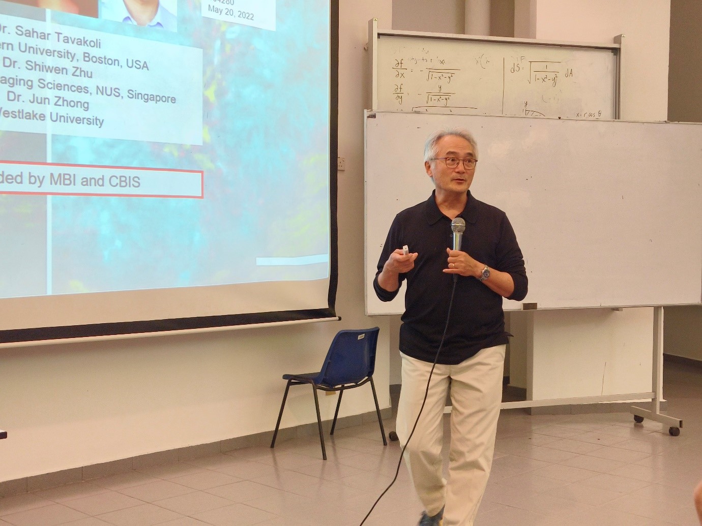
Prof Paul introducing some relevant research done by his students
During the lecture, Prof Paul clarified the importance of the intestines in consuming nutrients. He said, “Our intestines are able to absorb food efficiently because we have an enormous surface area lining in our intestines. The intestines consist of finger-like structures called villi and microvilli, which together cause the intestinal surface area to be able to expand from 0.5m2 to 500m2.” He explained the significance of zebrafish’s intestinal structure in intestinal cell research. He said, “Zebrafish’s intestines are composed of zig-zagging villar ridges, which is the intermediate evolutionary form between the simplest flat surface and the complex surface covered by villi.”
To elaborate on the topic, Prof Paul shared the results of his past research to show the translocating cells in a zebrafish’s intestines. By using EdU as the DNA marker, he found that most of the mitotic cells and stem cells are located in the pockets of the intestines. As a result, Prof Paul and his team started questioning the specific location of the stem cell in the bottom of the pockets. They used stem cell markers such as BMI1, Lrig1, and PRMT1 to identify the exact location. In the process, they discovered cell clusters in the pockets of the Zebrafish’s intestines. Prof Paul elucidated that the cell clusters are visually and functionally alike to crypts in the developed intestinal structure of other creatures. Hence, Prof Paul and his team concluded that the cell clusters are the functional and structural evolutionary precursors to crypts.
The lecture concluded with an insightful Q&A session between Prof Paul and UTAR staff and students.
Also present at the lecture were UTAR Faculty of Science (FSc) Dean Assoc Prof Dr Lim Tuck Meng, FSc Deputy Dean for Research and Development and Postgraduate Programmes Dr Phoon Lee Quen, NUS Department of Biological Sciences Emeritus Prof Dr Hew Choy Leong, UTAR staff and students.
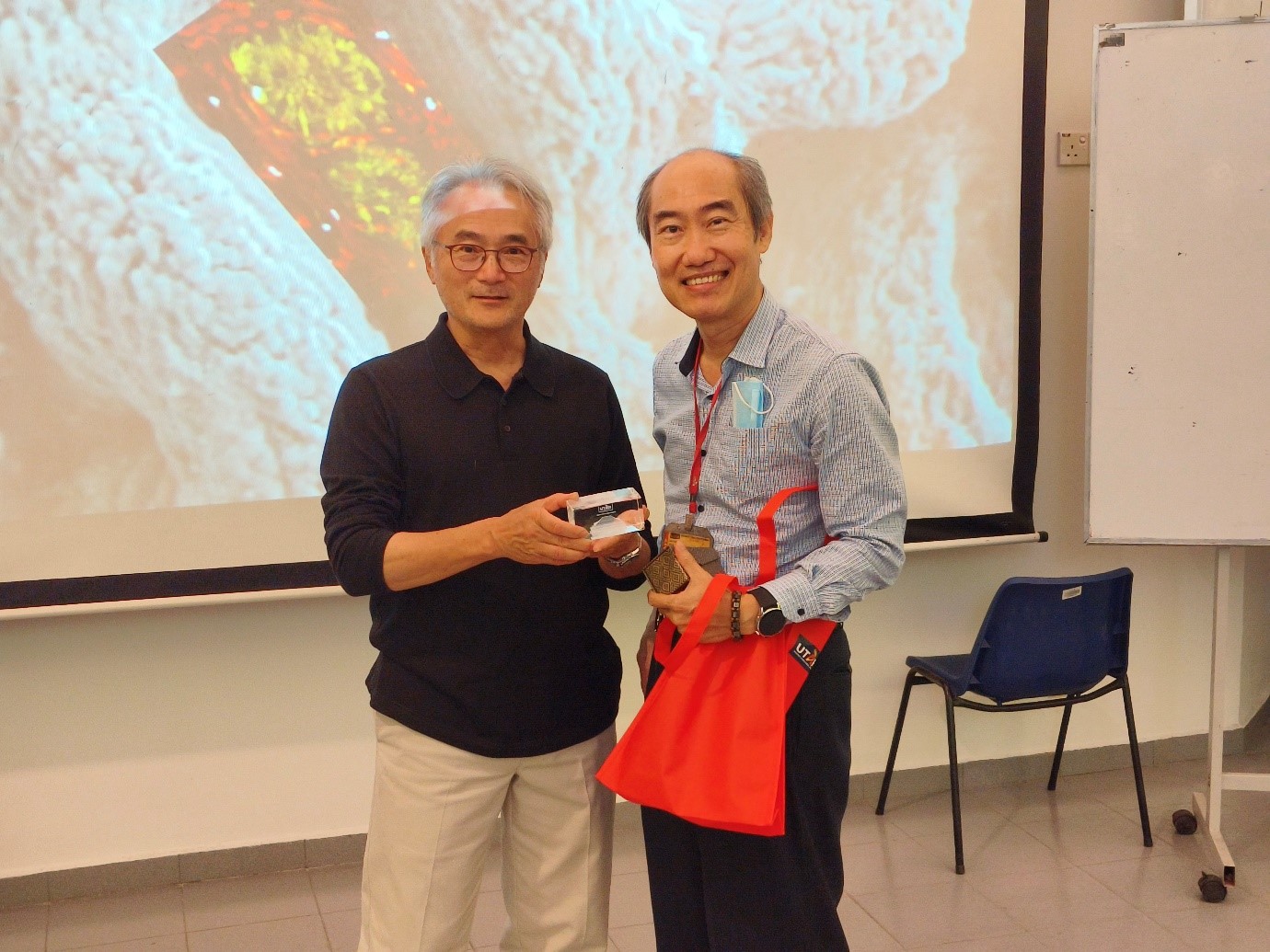
Dr Lim (right) presenting a token of appreciation to Prof Paul
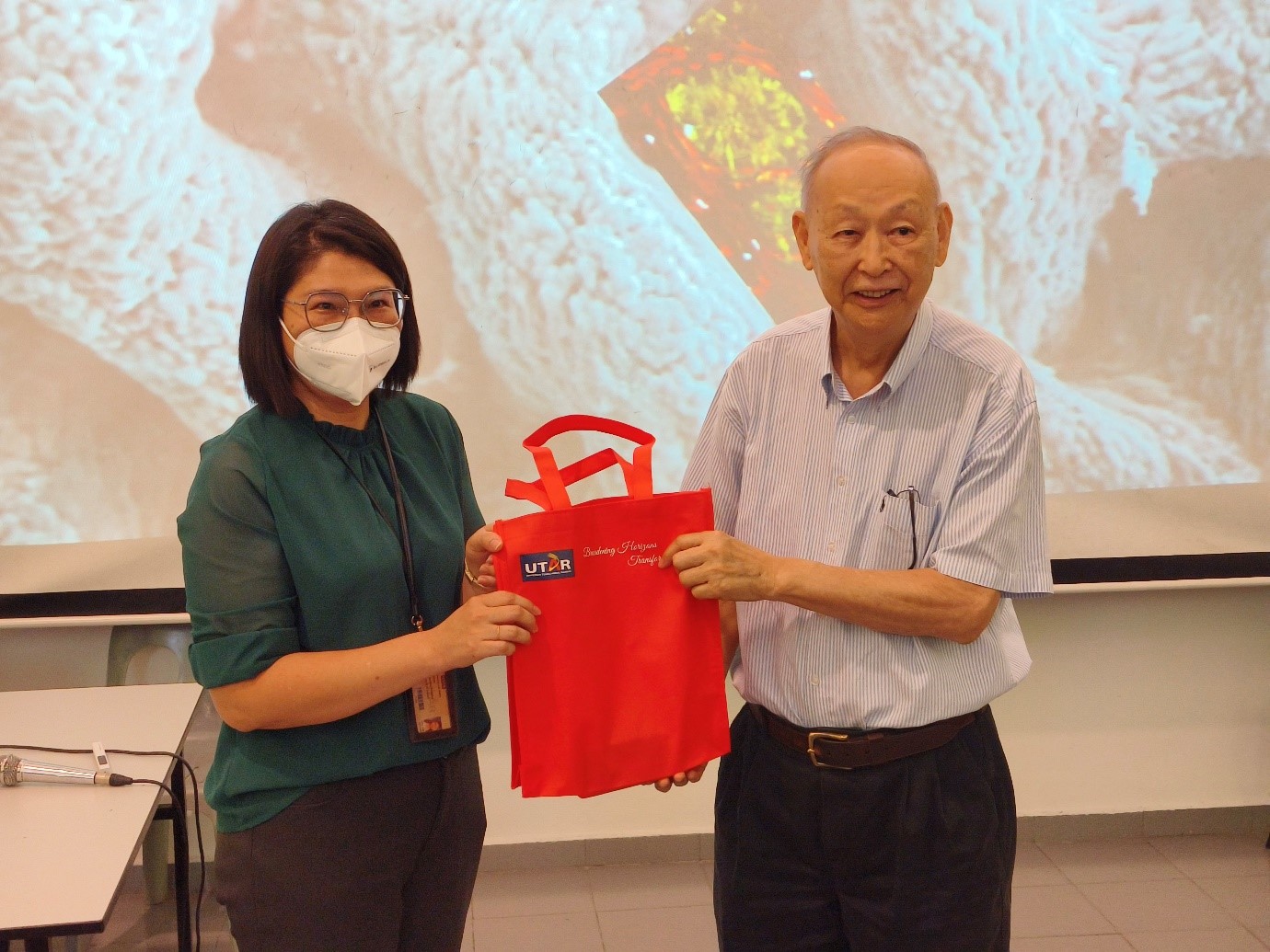
Dr Phoon (left) presenting a token of appreciation to Prof Hew
© 2023 UNIVERSITI TUNKU ABDUL RAHMAN DU012(A).
Wholly owned by UTAR Education Foundation (200201010564(578227-M)) LEGAL STATEMENT TERM OF USAGE PRIVACY NOTICE